Human brain theory
for the European Union’s Human Brain Project
ISBN 978-3-00-068559-0
15.The evolution of the basal ganglia system
Created on 12.02.2024 by Dr Andreas Malczan
The main task of new neuronal circuits is to acquire new signals.
The main task of the basal ganglia system initially consisted of obtaining differential images to recognise changes in signal strength through the multisensory analysis of moving objects. Only later, with the development of the cerebellum and signal divergence and the inclusion of the limbic system, did the basal ganglia make it possible to link signals from the past with signals from the present. This was also a prerequisite for linking cause and effect.
The author only came to this realisation last year; previously, he was of the opinion that the formation of differential images was the sole main objective of the basal ganglia system.
However, the development of a theory of language learning was only possible if there was a transition in the basal ganglia from difference mapping to the generation of excitatory past signals.
How is this to be understood? Let's take a simple cause: an animal eats something that is not digestible. While eating, it can learn the taste and odour of what it has eaten. The pontocerebellum serves this purpose. The cause can therefore be learnt.
A little later, the animal gets sick. The animal can also learn the associated signals. The effect is therefore also memorised.
However, if the animal cannot link the cause with the effect, it will eat the unpalatable food again and again, and it will always get sick afterwards. If the eaten food is even really toxic after repeated ingestion, a non-learning animal will ultimately die from it.
The animal must therefore be able to link two types of signal: Past signals with present signals. But there is a problem with this: past signals are no longer readily available. Their lifespan is limited, they are simply the past.
Therefore, the vertebrate nervous system had to undergo a further development in which the past signals were transported into the present so that they could be combined with present signals.
This was achieved by the early basal ganglia system. It sent the signals on a journey during which some time passed, and in the meantime the present had already begun.
In the early vertebrate brain, there were only analogue signals whose firing rate represented the strength of sensory signals. Thus, the firing rate of the odour receptors increased with the intensity of the odour. Similarly, the firing rate of muscle signals, for example from the tendon organs, increased with muscle tension. All signals were initially analogue.
Changes in signal strength could also indicate the approach or distance of objects that were smelled, felt or seen. The early striosome system of the basal ganglia made it possible to recognise changes in signal strength.
Even then, all sensory signals reached the sensory cortex in ascending order. The signals reached the motor side of the cortex via the axons of the class 3 neurones and descended again to ultimately activate the motor neurones of the trunk via the nucleus ruber. All modalities were more or less used to control movement with the aim of foraging and escaping from predators.
However, the descending cortex signals also reached other substructures, such as the various average nuclei of the brain system. Below the thalamus was the average system of the subthalamic nucleus. Not only the nucleus ruber, but also the dopaminergic median centre, known as the substantia nigra, was located in the initial level of the brain. The downward-projecting cortex axons docked on their way to these two mean value structures and supplied the subthalamic nucleus and the substantia nigra pars compacta with excitatory input. Averaging nuclei inevitably had to integrate all available signals into their averaging process. Among other things, they themselves served to control important life processes.
Now mean value nuclei almost always project back into the structures of origin. This usually serves to activate the sensory or motor areas so that, for example, foraging could be activated before the lack of energy became too great.
The substantia nigra pars compacta also projected back towards the cortex. Since it used the transmitter dopamine, there was a dopaminergic projection from it to the cortex.
Activation of the cortex neurons would have led to a neuronal build-up of the signal strength, as the cortex in turn projected back. Therefore, the dopaminergic axons docked onto the inhibitory interneurons of the cortex and excited them. However, their original task was to inhibit the neighbouring cortex neurons. This inhibitory effect has now been strengthened. In the course of evolution, these inhibitory interneurons in the cortex became independent and formed their own neuronal layer below the cortex neurons. This layer became the primitive striatum. The GABAergic neurones in this layer developed axon collaterals that projected downwards, just as the cortex neurones did. In the course of evolution, they reached the nucleus ruber. This is where the differential mapping described above could develop. The inhibitory signals from the primitive striatum overlapped with the incoming cortex signals, which were excitatory. However, the inhibitory signals had taken the longer diversions via the dopaminergic median nucleus and were time-delayed, i.e. past signals.
This gave rise to a new type of signal in the nucleus ruber, the class of time-sensitive differential signals. The inhibitory striatal neurons, which originally served to inhibit neighbours, slowly lost synaptic contact with the cortex neurons that had originally inhibited them. This meant that the primal striatum was able to function.
Nevertheless, there was a target that had not yet been reached. Although the output of the striatum was the past signal, this signal was inhibitory. An excitatory variant was required for a combination with the present signals. An excitatory past signal could converge with excitatory present signals on common output neurones and thus link the two in a meaningful way.
One possibility arose by applying the principle of including as many signals as possible in the averaging of mean value centres.
The inhibitory signals descending from the primitive striatum to the nucleus ruber passed directly by the subthalamic nucleus on their way. In the course of evolution, these signals also interacted with the subthalamic nucleus. They docked onto the inhibitory interneurones of the subthalamic nucleus. Just as the dopaminergic signals used to contact inhibitory interneurones in the cortex, the descending signals of the striatal neurones made synaptic contact with the inhibitory interneurones of the subthalamic nucleus and inhibited them.
However, as these neurones were actively excited by the neurones of the subthalamic nucleus, a signal inversion now occurred. The reason was the relative inhibition; the mean value signals of the subthalamic nucleus were too strong for total inhibition.
The ability of inhibitory neurons to make contact with the inhibitory interneurons of mean value nuclei and the subsequent formation of projection axons appears to be a basic ability in the basal ganglia system that occurs repeatedly and multiple times. We will encounter this ability several more times in the basal ganglia system.
The inhibitory interneurons of the subthalamic nucleus, which were inhibited by the GABAergic striatal neurons, became independent and formed their own nucleus and projection axons. The nucleus they formed was the globus pallidus, known as the globus pallidus interna because it would later split into an internal and an external subnucleus. Its projection neurones were GABAergic, i.e. inhibitory. They had not only received the mean value excitation from the subthalamic nucleus, but also the place markers. Therefore, these axons now travelled upwards again, where they were the first structure to reach the thalamus. However, the thalamus also already contained averaging neurones, as do most neurone structures. They were apparently also located approximately in the centre of the thalamus structure at that time. The thalamus thus had a kind of median area, a subnucleus with median excitation.
The ascending axons of the newly formed globus pallidus met these excitatory median neurones in the thalamus and docked onto them. These median neurones were strongly excited by the thalamic median, so the signals from the globus pallidus only inhibited them relatively. This corresponded to a signal inversion.
The thalamic median neurons contacted by the globus pallidus separated in the course of evolution and formed their own nucleus, which was located next to the previous thalamus and is also referred to as the thalamus by neurologists. This new thalamic nucleus was given the name nucleus centromedianus by neurologists. Its neurons took over the location markers of the original median neurons from which they had emerged and therefore travelled upwards to the cortex.
The signals of the nucleus centromedianus were the doubly inverted, glutamatergic and time-delayed cortex signals. The substantia nigra pars compacta was responsible for the time delay. The striatal neurones were responsible for switching to GABA. The first inversion took place in the globus pallidus, the output was also inhibitory. The second inversion took place in the nucleus centromedianus, where thalamic median signals were relatively inhibited. The output was now excitatory and used glutamate as a transmitter. It represented the time-delayed past signal, which was now available in the present due to the time delay and could be combined with present signals.
Note:
The striosomes of the striatum are a descendant of the inhibitory interneurones of the cortex. The globus pallidus interna is a descendant of the inhibitory interneurones of the subthalamic nucleus. The centromedian nucleus of the thalamus is a descendant of the excitatory median neurones of the median neurones of the thalamus.
Since we have always considered those striatal neurones that are excited by dopamine in the previous analysis, these are the striosomes of the striatum. Therefore, the following applies:
Output of the striosome system
The output of the striosome system, which reaches the cortex again, represents excitatory past signals that have been transformed into the present by a time delay on their dopaminergic diversions. They can now be combined with present signals. These signals are all fire-rate coded, i.e. they represent signals from the analogue brain system.
But evolution did not stand still. Signalling diverged step by step, starting in the olivary nucleus and later also in other nuclei and in the cortex. This gave rise to the class of extreme value-coded signals. The basal ganglia system also had to find a solution for this class of signals in order to create both a differential circuit for recognising movement and an excitatory variant of the excitatory past signals. The part of the striatum involved in this task was the matrix. It processed the extreme value coded signals of the brain.
The extreme-value coded signals were generated from the analogue signals by signal divergence. The following diagram from the author's monograph "Brain Theory of Vertebrates" serves to illustrate signal divergence in the nucleus olivaris:
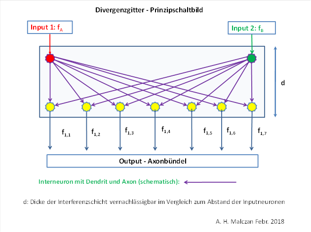
It can be seen that the signal divergence creates an entire signal vector of extreme value-coded signals from the original two (analogue) signals, which also contains the two original signals. Minimum coding occurs in the olivary nucleus with linear divergence.
These newly added signals are processed in the basal ganglia system by the matrix system, which works somewhat differently from the striosome system.
Initially, the minimum-coded signals rise from the nucleus olivaris as climbing fibres to the cerebellum, where they strongly excite the Purkinje cells. The Purkinje cells inhibit the permanently excited cerebellar nuclei, which receive their permanent excitation from the median nucleus of the formatio reticularis. The relative inhibition of this permanent excitation leads to the first inversion of the signals. The spinocerebellum serves as an inversion circuit.
Once inverted, the signals are now maximum-coded and reach the cortex in ascending order. The cortex region is located in the frontal cortex, which receives the cerebellum output.
The cortex in turn projects back into the dopaminergic median nucleus, the substantia nigra pars compacta. There, the cortex signals are switched to dopamine and return to the striatum.
Here, however, the mode of operation changes. GABAergic neurones are contacted again in the striatum. These were originally also used for neighbour inhibition. For this purpose, each interneuron contacted a large number of cortex neurons and received their excitation; the axon of the interneuron inhibited exactly one cortex neuron. Thus, the environment of this neuron had an inhibitory effect. If the environment was relatively strongly excited, this inhibitory interneuron was also strongly excited.
When a returning dopaminergic axon docked onto it and inhibited it, a signal inversion took place. The maximum-coded cortex output was inverted by this signal inversion.
Thus, the first signal inversion of the maximum-coded cortex signals of the frontal cortex already took place in the matrix system within the striatum. The inhibitory interneurons acquired the ability to form their own projection axon, which projected downwards. The output of the matrix was inhibitory and minimum-coded.
Just like the striosome axons, the matrix axons travelled towards the subthalamic nucleus and contacted inhibitory interneurons there, just as the striosome axons did. These interneurones also separated, formed projection axons and thus also became a component of the already existing globus pallidus. However, these neurones also separated spatially and formed their own segment in the globus pallidus. The original segment formed the globus pallidus interna, while the new segment became the globus pallidus externa, positioned above between the striatum and globus pallidus interna. This created a new structure in the basal ganglia system.
The output of the globus pallidus externa was inhibitory, the transmitter was GABA, the signals were now inverted again, i.e. maximum-coded. Technically, they differed from the minimum-coded signals of the globus pallidus interna only in the type of coding. This nucleus was exactly the structure that still had to be passed through. Therefore, these signals were treated analogue.
The axons of the new globus pallidus externa travelled in the direction of the globus pallidus interna and also generated GABAergic projection neurons there, which in turn received excitatory mean input from the subthalamic nucleus and thus inverted the input of the globus pallidus externa. This meant that the output of the matrix system from the globus pallidus interna was in turn inhibitory and minimum-coded. The signals obtained in this way travelled further in the direction of the thalamus, but had to travel to those thalamic structures that received the original, maximum-coded signals from the cerebellum. In these target areas, too, there was increased averaging by average neurones. Here, the basal ganglia signals of the matrix system, which came from the globus pallidus interna, docked and made synaptic contact with such averaging neurones. The relative inhibition of the median signals corresponded to a new signal inversion, their output signals were now excitatory and again maximally encoded and corresponded to the time-delayed cortex signals from which they had emerged. However, they represented a new class of signals: they were excitatory past signals that were available to the present. They had reached the present via the dopaminergic substantia nigra pars compacta.
The associated output neurons separated spatially from the thalamic median neurons and formed new thalamic structures. This is how the anterior ventral nucleus (VA) and the lateral ventral nuclei (VL) emerged from the thalamus. They were the result of the evolutionary development of the basal ganglia system under the influence of signalling divergence in the spinocerebellum.
Now the past signals were available in their exciting form and could be combined with the present signals.
We summarise the functioning of the basal ganglia in the following overview.
|
|
Striosomes |
Matrix |
|
Cortex signals |
analogue, exciting Glutamate |
maximumcoded, exciting, Glutamate |
|
Substantia nigra pars compacta |
Dopamine, exciting |
Dopamine, inhibiting |
|
Striatum |
Gaba, rate of fire is maintained |
Gaba, inversion neurones, Excitation through NST Inhibition by dopamine Output inhibiting, minimum coded |
|
Globus pallidus interna |
is traversed without impact |
Gaba, Inversion neurones, Excitation through NST Inhibition by GABA Output inhibiting, maximumcoded |
|
Globus pallidus externa |
Gaba, firing rate is inverted, Inversion neurones, Excitation through NST Inhibition by GABA Output inhibiting |
Gaba, Inversion neurones, Excitation through NST Inhibition by GABA Output inhibiting, minimumcoded |
|
Thalamus, inhibiting variant |
A copy of the thalamic signals is inhibited by the globus pallidus externa in a point-to-point mapping, Time-sensitive difference mapping |
A copy of the thalamic signals is inhibited by the globus pallidus externa in a point-to-point mapping, Time-sensitive difference mapping |
|
Thalamus, median nucleus |
glutamatergic inversion neurones, Excitation from the mean value core, Inhibition by globus pallidus externa, Output exciting, Time-shifted cortex signals (excitatory past signals) |
glutamatergic inversion neurones, Excitation from the mean value core, Inhibition by globus pallidus externa, Output exciting, Time-shifted cortex signals (excitatory past signals) |
As can be seen in the table, both the formation of the time-sensitive difference mapping for motion detection in the basal ganglia system and the formation of the excitatory, time-shifted past signals are postulated. While the author has recognised and described the formation of the time-sensitive difference mapping in the basal ganglia system in previous publications, the formation of the excitatory past signals was only recognised by him in the last half of 2023. The reason for the maturation of this insight arose because the learning of language by means of the pontocerebellum, which is capable of learning, could not be explained otherwise.
The author still believes that the brain basically processes, stores and combines signals. The transfer of past signals into the present makes this possible. And the easiest way to transfer signals from the past to the present is through echo formation. Anyone can try this out in the mountains: A loud call travels at the speed of sound and is reflected by the mountain walls. As it propagates, the clock continues to run. This means that the sound reaches the listener at a much later point in time than it was generated, i.e. it is a signal from the (recently elapsed) past.
The basal ganglia utilise the lower propagation speed and the longer signal path via the dopaminergic median nucleus to achieve such a time delay of the signal.
Now we have to deal with the size of the time delay. This is so small that it now requires further subsystems of the brain.
The time delay in the basal ganglia system is well below the second range; the author estimates it at around 25 to 50 milliseconds. This means that the echo is still active about 25 to 50 milliseconds after the end of the output signal. This time is too short to cause any learning processes in the pontocerebellum by means of LTP and LTD.
The brain therefore needs a way to significantly extend this signal duration so that it is at least one second or more. The longer the echo duration of a signal, the greater the time span with which it can be combined with other, later activated signals.
A structure that had existed in the vertebrate brain since time immemorial could contribute to solving this problem. This structure was the Papez circle.
Every signal that is fed into the Papez circuit rotates continuously in a closed loop with the following intermediate stations:
·Hippocampus
·Corpus mammilare
·Anterior thalamic nucleus
·Cingulate gyrus
·Area entorhinalis
·Parahippocampal gyrus
·Tractus perforans
·Hippocampus
The rotation of signals in the limbic loop, as the Papez circuit is also known, could theoretically continue forever. In practice, however, there is access via stop signals that can excite inhibitory interneurons at various points in the loop, which in turn stop the signalling rotation. The internal clock in the brain in the suprachiasmatic nucleus can deliver such stop signals to the limbic system. But other regions can also do this. In particular, neuronal competition can occur between the limbic signals, so that one signal can suppress other signals. We will encounter this in the next chapter.
The limbic system can significantly prolong the rather short signal echoes of the basal ganglia system, so that their lifespan can extend far beyond the seconds or even minutes range. All that is required is that these signals are fed into the limbic system as input. If they rotate in loops there, they can reach the pontocerebellum as output via the bridge nuclei and the Kleter fibres with each signal circulation. Now the pontocerebellum can use these past signals to be combined with present signals. The author will explain how this works in the next chapter. This chapter explains which algorithms in the brain in which subsystems enable spoken language to be learnt. The learning of written language will probably be dealt with in a later chapter.
Monografie von Dr. rer. nat. Andreas Heinrich Malczan